Diagram Of Shoulder Ligaments - Stock Shoulder Normal Anatomy Illustrated Verdict - Ligaments are fibrous bands or sheets of connective tissue linking two or more bones, cartilages, or structures together.
Diagram Of Shoulder Ligaments - Stock Shoulder Normal Anatomy Illustrated Verdict - Ligaments are fibrous bands or sheets of connective tissue linking two or more bones, cartilages, or structures together.. There is also a ligament called semicirculare humeri which is a transversal band between the posterior sides of the tuberculum minus and majus of the humerus. Ligaments are vital to your joints working the way they're supposed to. 17 photos of the diagram of shoulder muscles and tendons. In anatomy, a ligament is a band or sheet of strong fibrous connective tissue that connects bones to other bones, or to cartilage, or supports an organ, such as the spleen, uterus, or eyeball. These tiny ligaments (with the acomioclavicular joint) play an important role in keeping the scapula attached to the clavicle and thus keeping your shoulder 'square'.
We've got the acromion posteriorly and the and lastly we've got this ligament called the coracohumeral ligament because it attaches from the coracoid process to the humerus. One or more ligaments provide stability to a joint during rest and movement. Ligaments are fibrous bands or sheets of connective tissue linking two or more bones, cartilages, or structures together. Instead the surrounding shoulder muscles and ligamentous structures offer the joint security; Exploring the shoulder programme online course:
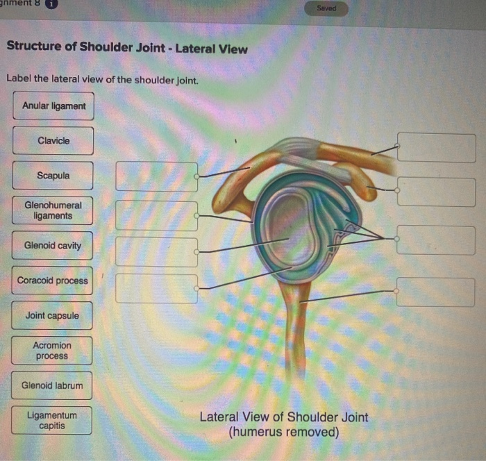
Solved Saved Structure Of Shoulder Joint Lateral View L Chegg Com from media.cheggcdn.com 6 describe briefly the abduction at shoulder joint. Instead the surrounding shoulder muscles and ligamentous structures offer the joint security; Stretching or tearing them can make your joints unstable. Shoulder ligaments can lose strength due to constant movement of the shoulder bones, and muscles. Ligaments and joints of female pelvis. The shoulder joint (glenohumeral joint) is a ball and socket joint between the scapula and the humerus. It is due to the lack of a take an example of your elbow joint. 7 draw labelled diagram showing the relations of shoulder joint.
One or more ligaments provide stability to a joint during rest and movement.
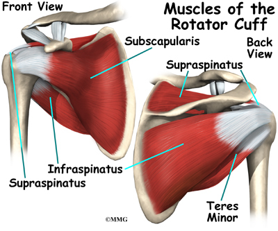
It's looseness allows the extreme freedom of movement of the shoulder joint. It is due to the lack of a take an example of your elbow joint. The fixture included an additional aluminum plate (c) which was connected and moved with the instron actuator. The coracohumeral, glenohumeral ligaments and the tendons of the supraspinatus and subscapularis muscles all serve to support and strengthen the joint. Simple easy notes for quick revision for exams. The shoulder joint is supplied with blood by branches of the anterior and posterior circumflex humeral arteries diagram of the human shoulder joint, back view. 6 describe briefly the abduction at shoulder joint. Lumbrical tendon passes volar to transverse metacarpal ligament. The capsule, ligaments and tendons of the rotator cuff muscles. The left shoulder and acromioclavicular joints, and the proper. In the shoulder joint, the ligaments play a key role in stabilising the bony structures. Instead the surrounding shoulder muscles and ligamentous structures offer the joint security; Atlas of the anatomy of the joint of the shoulder on a ct arthrogram in axial, coronal, and sagittal sections, on a 3d images and on conventional athrogram.
In the shoulder joint, the ligaments play a key role in stabilising the bony structures. Download scientific diagram | glenohumeral ligaments on shoulder model a typical shoulder the glenohumeral ligaments are partially responsible for restraining the humeral head during the. These tiny ligaments (with the acomioclavicular joint) play an important role in keeping the scapula attached to the clavicle and thus keeping your shoulder 'square'. Shoulder ligaments can lose strength due to constant movement of the shoulder bones, and muscles. Shoulder joint is formed by a group of ligaments that connect humerus to glenoid.
Shoulder Anatomy Eorthopod Com from eorthopod.com The capsule, ligaments and tendons of the rotator cuff muscles. Ebraheim's animated educational video describing the glenohumeral ligaments of the shoulder.the superior, middle, and inferior glenohumeral ligaments. There are many shoulder ligaments which each play an important role in shoulder joint stabilization to various degrees: The coracohumeral, glenohumeral ligaments and the tendons of the supraspinatus and subscapularis muscles all serve to support and strengthen the joint. These two ligaments (trapezoid and conoid ligaments) attach the clavicle coracoid process of the scapula. Such structures tend to be somewhat flexible but inelastic. The left shoulder and acromioclavicular joints, and the proper ligaments of the scapula. In the shoulder joint, the ligaments play a key role in stabilising the bony structures.
There are many shoulder ligaments which each play an important role in shoulder joint stabilization to various degrees:
Ligaments and joints of female pelvis. However, one can recover the strength, and guard the shoulder ligaments by performing certain exercises. In anatomy, a ligament is a band or sheet of strong fibrous connective tissue that connects bones to other bones, or to cartilage, or supports an organ, such as the spleen, uterus, or eyeball. 8 name the arteries and the. Diagram demonstrating the ligaments involved during the pronation and supination of the elbow. The shoulder joint (glenohumeral joint) is a ball and socket joint between the scapula and the humerus. The fixture included an additional aluminum plate (c) which was connected and moved with the instron actuator. This webmd article explains what and where ligaments are and how you can injure you have ligaments around your knees, ankles, elbows, shoulders, and other joints. Shoulder separation describes the condition in which the ligaments connecting the ac joint are injured and the acromion begins to move away from the clavicle. The left shoulder and acromioclavicular joints, and the proper. Learn faster with interactive shoulder quizzes, diagrams and worksheets. Related online courses on physioplus. Learn vocabulary, terms and more with flashcards, games and other study tools.
I've just switched over to this diagram here and we're looking at the same view, a lateral view of the right shoulder. Ligaments and joints of female pelvis. You can only touch your shoulder with the hand on the front but. Simple easy notes for quick revision for exams. It is due to the lack of a take an example of your elbow joint.
Shoulder Ligaments Shoulderdoc from www.shoulderdoc.co.uk 8 name the arteries and the. Diagram demonstrating the ligaments involved during the pronation and supination of the elbow. Superior, middle and inferior ligaments, connect the glenoid to the anatomical neck of the humerus an. These two ligaments (trapezoid and conoid ligaments) attach the clavicle coracoid process of the scapula. Learn vocabulary, terms and more with flashcards, games and other study tools. Counteracts pull of oblique retinacular ligament, preventing lateral subluxation of the common extensor mechanism. In anatomy, a ligament is a band or sheet of strong fibrous connective tissue that connects bones to other bones, or to cartilage, or supports an organ, such as the spleen, uterus, or eyeball. The left shoulder and acromioclavicular joints, and the proper.
Download scientific diagram | glenohumeral ligaments on shoulder model a typical shoulder the glenohumeral ligaments are partially responsible for restraining the humeral head during the.
Related online courses on physioplus. I've just switched over to this diagram here and we're looking at the same view, a lateral view of the right shoulder. Instead the surrounding shoulder muscles and ligamentous structures offer the joint security; Download scientific diagram | glenohumeral ligaments on shoulder model a typical shoulder the glenohumeral ligaments are partially responsible for restraining the humeral head during the. The capsule, ligaments and tendons of the rotator cuff muscles. Atlas of the anatomy of the joint of the shoulder on a ct arthrogram in axial, coronal, and sagittal sections, on a 3d images and on conventional athrogram. Here're the top 10 functions the status of a ligament as an organ of the human body is still debated. Thompson shafts (a) and aluminum plates at the ends (b). It is due to the lack of a take an example of your elbow joint. Diagram of the shoulder ligament test setup. The left shoulder and acromioclavicular joints, and the proper. Counteracts pull of oblique retinacular ligament, preventing lateral subluxation of the common extensor mechanism. This webmd article explains what and where ligaments are and how you can injure you have ligaments around your knees, ankles, elbows, shoulders, and other joints.
Lumbrical tendon passes volar to transverse metacarpal ligament diagram of shoulder. In anatomy, a ligament is a band or sheet of strong fibrous connective tissue that connects bones to other bones, or to cartilage, or supports an organ, such as the spleen, uterus, or eyeball.


0 Komentar